Back Muscles Anatomy : Muscles Of Back Simplified Epomedicine / Anterior rami of spinal nerve innervate them.. Muscles of the back can be divided into superficial, intermediate, and deep group. The upper back is a complex area containing a number of muscles that perform various actions on the scapulae (shoulder blades) and humerus. We hope this picture anatomy of back muscles diagram can help you study and research. All these muscles are therefore associated with movements of the upper limb. Back muscles, functions and exercises:
The former two groups, superficial and intermediate, are referred to as the extrinsic back muscles. The back muscles are anatomically layered into superficial (extrinsic) and deep (intrinsic) muscles. They start at the top of the neck and go down to the tailbone. For more anatomy content please follow us and visit our website: All these muscles are therefore associated with movements of the upper limb.
Back Muscles Stock Photos And Images 123rf from us.123rf.com Anatomy of back muscles your back consists of three distinct layers of muscles, namely the superficial layer, the intermediate layer, and the deep layer. There are three major groups of back muscles:. These structures work together to support the body, enable a range of movements, and send messages from the brain to the. On this page, you'll learn about each of these muscles, their locations and functional anatomy. We hope this picture anatomy of back muscles diagram can help you study and research. The multifidus, a long muscle that travels nearly the entire length of the back.it helps to stabilize and rotate the lower back, and additionally takes some. Muscle origin insertion action innervation artery notes; Anterior rami of spinal nerve innervate them.
Memorize all the muscle facts with the help of muscle cheat sheets.
Extends and laterally bends the neck and head, rotates head to the same side: The intrinsic back muscles are found deeper to the extrinsic muscles, separated from them by the thoracolumbar fascia. These muscles give height and breadth to back development. What are the lower back muscles and their anatomy? Both the deltoid and the trapezius are firmly attached to the spine of the scapula. Artery) p.134 accessory nerve p. Back muscles, functions and exercises: The deltoid, teres major, teres minor, infraspinatus, supraspinatus (not shown) and subscapularis muscles (not shown) all extend from the scapula to the humerus and act on the shoulder joint. The former two groups, superficial and intermediate, are referred to as the extrinsic back muscles. These muscles work together to move the scapula anteriorly and laterally during pushing, throwing, or punching motions. Superficial back muscles, intermediate back muscles and intrinsic back muscles.the intrinsic muscles are named as such because their embryological development begins in the back, oppose to the superficial and intermediate back muscles which develop elsewhere and are therefore classed as extrinsic muscles. The latter group is the intrinsic muscle group. The upper back is a complex area containing a number of muscles that perform various actions on the scapulae (shoulder blades) and humerus.
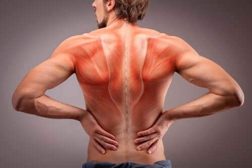
The superficial back muscles are situated underneath the skin and superficial fascia. In the upper back region, the trapezius, rhomboid major, and levator scapulae muscles anchor the scapula and clavicle to the spines of several vertebrae and the occipital bone of the skull. The multifidus, a long muscle that travels nearly the entire length of the back.it helps to stabilize and rotate the lower back, and additionally takes some. The latter group is the intrinsic muscle group. Muscles of the back can be divided into superficial, intermediate, and deep group.
What Is The Anatomy Of Back Muscles Quora from qph.fs.quoracdn.net The extensor muscles are attached to back of the spine and enable standing and lifting objects. Balance the weight of your head on top of your spine evenly distribute weights from your upper body into the lower extremities Both the deltoid and the trapezius are firmly attached to the spine of the scapula. The back muscles are anatomically layered into superficial (extrinsic) and deep (intrinsic) muscles. Muscles that act on the back. Anatomy of back muscles your back consists of three distinct layers of muscles, namely the superficial layer, the intermediate layer, and the deep layer. Anterior rami of spinal nerve innervate them. The multifidus, a long muscle that travels nearly the entire length of the back.it helps to stabilize and rotate the lower back, and additionally takes some.
The upper back is a complex area containing a number of muscles that perform various actions on the scapulae (shoulder blades) and humerus.
Back muscles the muscles of the back are a group of strong, paired muscles that lie on the posterior aspect of the trunk. Attached to the shoulder girdle intermediate: The multifidus, a long muscle that travels nearly the entire length of the back.it helps to stabilize and rotate the lower back, and additionally takes some. The muscles, bones, ligaments, and tendons in the back can all be injured and cause back pain. The back consists of the spine, spinal cord, muscles, ligaments, and nerves. Muscle or ligament strains can occur from repeated use of the muscles, or from improperly or awkwardly lifting heavy objects. The deep muscles develop in the back called intrinsic muscles. For more anatomy content please follow us and visit our website: Back pain is common and might be caused by a problem with a muscle. Attached to the posterior thorax; Extends and laterally bends the neck and head, rotates head to the same side: We hope this picture anatomy of back muscles diagram can help you study and research. This curve, called lordosis, helps to:
The superficial back muscles are situated underneath the skin and superficial fascia. 1 your spine in this region has a natural inward curve. The muscles, bones, ligaments, and tendons in the back can all be injured and cause back pain. There are three major groups of back muscles:. Deep muscles of the lower back include:
The Anatomy Of The Back Muscles Step To Health from steptohealth.com We hope this picture anatomy of back muscles diagram can help you study and research. Balance the weight of your head on top of your spine evenly distribute weights from your upper body into the lower extremities Anatomy of the back muscles the latissimus dorsi muscles (also known as the lats) are the largest muscles of the back. The intrinsic back muscles are found deeper to the extrinsic muscles, separated from them by the thoracolumbar fascia. Back muscles, functions and exercises: Leaning back to straight vertical and all points in between. Back muscles the muscles of the back are a group of strong, paired muscles that lie on the posterior aspect of the trunk. For more anatomy content please follow us and visit our website:
These muscles give height and breadth to back development.
The superficial back muscles are situated underneath the skin and superficial fascia. Memorize all the muscle facts with the help of muscle cheat sheets. Muscle origin insertion action innervation artery notes; The back muscles are anatomically layered into superficial (extrinsic) and deep (intrinsic) muscles. Anatomynote.com found anatomy of back muscles diagram from plenty of anatomical pictures on the internet. Your lower back (lumbar spine) is the anatomic region between your lowest rib and the upper part of the buttock. The muscles of the lower back, including the erector spinae and quadratus lumborum muscles, contract to extend and laterally bend the vertebral column. This is a tutorial to quickly s. The extrinsic back muscles are located in the back, but act to produce movements of the shoulder and assist respiration. Balance the weight of your head on top of your spine evenly distribute weights from your upper body into the lower extremities Artery) p.134 accessory nerve p. Understanding lower back anatomy is key to understanding the root of lower back and hip pain. Attached to the shoulder girdle intermediate:
Back Muscles Anatomy : Muscles Of Back Simplified Epomedicine / Anterior rami of spinal nerve innervate them.. There are any Back Muscles Anatomy : Muscles Of Back Simplified Epomedicine / Anterior rami of spinal nerve innervate them. in here.